
Orthopedics (Bones, Joints & Spine): Individualized Homeopathic Care in Dubai
Comprehensive Homeopathic Care for Bone, Joint, Spine & Musculoskeletal Disorders in Dubai
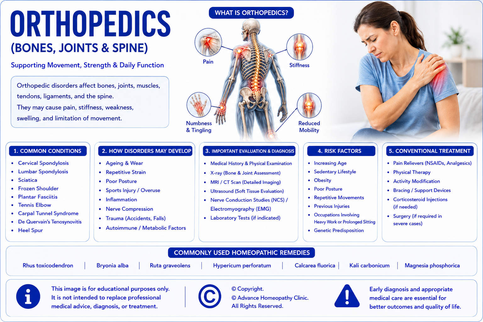
The musculoskeletal system provides the body with strength, stability, movement, and protection. Healthy bones, joints, muscles, tendons, ligaments, and the spine work together to allow everyday activities such as walking, climbing stairs, lifting objects, exercising, maintaining posture, and performing routine daily tasks. Disorders affecting these structures can lead to pain, stiffness, swelling, reduced flexibility, muscle weakness, numbness, and limitations in mobility that may significantly affect work, sports, and overall quality of life.
Orthopedic disorders may develop gradually as part of the natural ageing process, result from repetitive strain, poor posture, sports injuries, occupational overuse, inflammation, nerve compression, or underlying metabolic and autoimmune diseases. While some conditions improve with conservative treatment, others require long-term medical care, rehabilitation, or occasionally surgical intervention.
At Advance Homeopathy Clinic Dubai, we provide individualized homeopathic care for patients with a variety of orthopedic and musculoskeletal conditions, including Cervical Spondylosis, Lumbar Spondylosis, Sciatica, Frozen Shoulder (Adhesive Capsulitis), Plantar Fasciitis, Tennis Elbow (Lateral Epicondylitis), Carpal Tunnel Syndrome, De Quervain's Tenosynovitis, and Calcaneal Spur (Heel Spur). Homeopathic treatment aims to support the individual's overall health by considering the complete symptom picture, constitutional characteristics, medical history, lifestyle, and overall well-being rather than focusing solely on the orthopedic diagnosis.
Homeopathy is offered as a complementary approach alongside conventional orthopedic care. Our goal is to support symptom management, improve mobility, enhance quality of life, and promote overall well-being while encouraging patients to continue appropriate medical investigations, prescribed medications, physiotherapy, rehabilitation programmes, orthopaedic supports, and regular follow-up with their orthopaedic surgeon, spine specialist, sports medicine physician, or other healthcare professionals whenever required.
Understanding Orthopedic Disorders (Bones, Joints & Spine)
The musculoskeletal system consists of bones, joints, cartilage, muscles, tendons, ligaments, fascia, and the spine, all of which work together to provide support, movement, flexibility, and stability. Healthy joints allow smooth movement, while muscles generate force, tendons connect muscles to bones, ligaments stabilize joints, and the spine protects the spinal cord while allowing normal body movement.
Orthopedic disorders may develop through several different mechanisms, including:
- Age-related degeneration of joints and intervertebral discs
- Tendon and ligament overuse injuries
- Repetitive strain and occupational stress
- Sports-related injuries
- Inflammation affecting joints or soft tissues
- Nerve compression syndromes
- Muscle imbalance and poor posture
- Trauma or fractures
- Autoimmune and inflammatory disorders
- Metabolic conditions affecting bones and joints
- Congenital or inherited musculoskeletal disorders
Some orthopedic conditions develop suddenly following injury, while others progress gradually over many years. Early diagnosis is often essential because timely treatment may help reduce pain, preserve mobility, prevent permanent disability, improve physical function, and enhance long-term quality of life. Modern orthopedic care combines detailed clinical examination with imaging and other diagnostic investigations to establish an accurate diagnosis and guide evidence-based treatment.
How Homeopathy May Support Orthopedic Disorders
Homeopathy is an individualized system of medicine in which treatment is selected according to the patient's unique symptom pattern rather than the orthopedic diagnosis alone. During consultation, the homeopathic physician evaluates musculoskeletal complaints together with constitutional characteristics, medical history, emotional well-being, occupational demands, lifestyle, associated medical conditions, physical activity, and the overall pattern of symptoms before selecting an appropriate remedy.
Depending on the individual's clinical presentation, homeopathic treatment may be considered as a complementary approach for patients experiencing:
- Chronic neck pain and cervical spondylosis
- Lower back pain and lumbar spondylosis
- Sciatica with radiating leg pain
- Frozen shoulder with restricted movement
- Heel pain due to plantar fasciitis or calcaneal spur
- Tennis elbow and repetitive strain injuries
- Carpal tunnel syndrome
- De Quervain's tenosynovitis
- Tendon and ligament disorders
- Joint stiffness and reduced mobility
- Musculoskeletal pain affecting daily activities
Homeopathy complements conventional orthopedic care and should not replace appropriate medical investigations, prescription medications, physiotherapy, rehabilitation programmes, orthopaedic supports, image-guided procedures, or surgical treatment whenever these are medically indicated.
1. Cervical Spondylosis (Neck Arthritis)
What is Cervical Spondylosis?
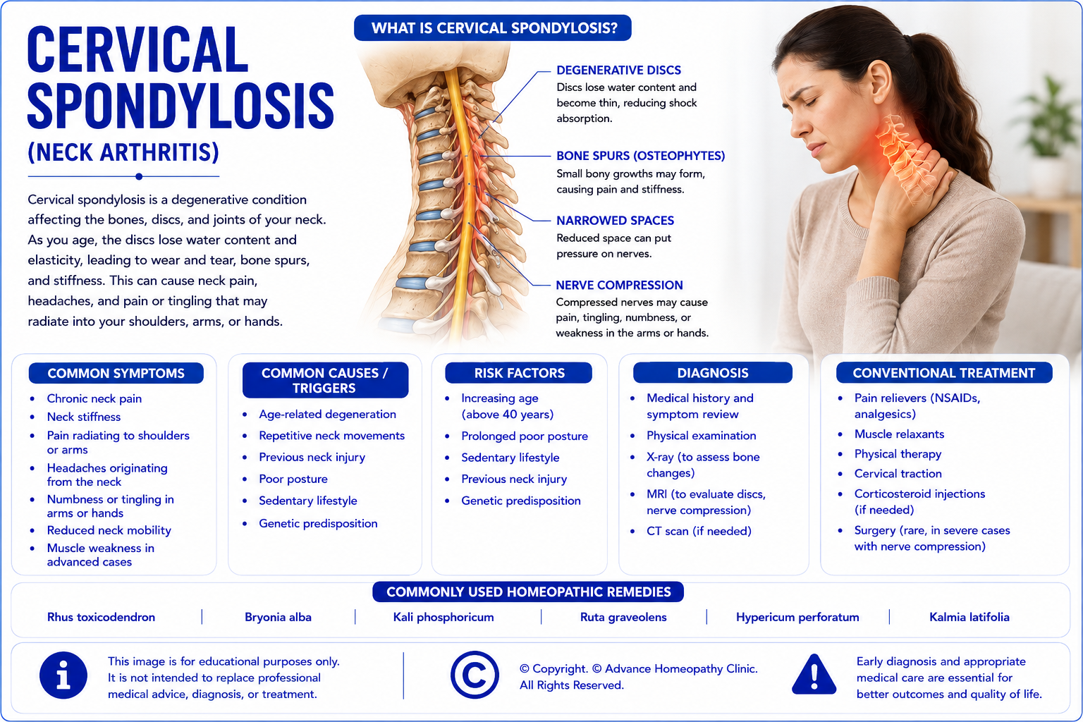
Cervical spondylosis, commonly known as neck arthritis, is a degenerative condition affecting the bones, intervertebral discs, joints, ligaments, and other supporting structures of the cervical spine (neck). As part of the natural ageing process, the spinal discs gradually lose water content and elasticity, leading to wear and tear of the cervical spine. Over time, these changes may result in narrowing of the spaces between the vertebrae, formation of bone spurs (osteophytes), stiffness, and reduced neck mobility.
In some individuals, these degenerative changes may compress nearby nerve roots or the spinal cord, causing pain that radiates into the shoulders or arms, numbness, tingling, muscle weakness, or difficulty with hand movements. Many people have age-related changes visible on imaging without significant symptoms, while others experience persistent pain and functional limitations that affect work, driving, sleep, and daily activities.
Common Signs & Symptoms of Cervical Spondylosis
Patients with cervical spondylosis may experience one or more of the following symptoms:
- Chronic neck pain
- Neck stiffness, especially after periods of inactivity
- Pain radiating to the shoulders, arms, or upper back
- Headaches originating from the neck (cervicogenic headaches)
- Reduced range of neck movement
- Numbness or tingling in the arms or hands
- Weakness of the upper limbs in some patients
- Muscle spasm around the neck and shoulders
- Pain that worsens after prolonged computer work or poor posture
Risk Factors for Cervical Spondylosis
Several factors may increase the likelihood of developing cervical spondylosis, including:
- Advancing age
- Degeneration of cervical intervertebral discs
- Previous neck injury or trauma
- Repetitive neck movements
- Poor posture and prolonged desk work
- Occupations requiring sustained neck positions
- Sedentary lifestyle
- Smoking
- Genetic predisposition
Conventional Management of Cervical Spondylosis
The treatment of cervical spondylosis depends on the severity of symptoms, neurological findings, and imaging results. Conservative treatment is effective for many patients and aims to reduce pain, improve neck mobility, strengthen supporting muscles, and maintain daily function.
Conventional management may include:
- Activity modification and ergonomic correction
- Physiotherapy and supervised exercise programmes
- Postural training
- Pain-relieving medications
- Non-steroidal anti-inflammatory drugs (NSAIDs)
- Muscle relaxants when indicated
- Cervical support devices for selected patients
- Image-guided spinal injections in appropriate cases
- Surgical decompression or spinal stabilisation for significant nerve or spinal cord compression
Homeopathic Medicines Commonly Considered for Cervical Spondylosis
Some homeopathic medicines that may be considered by experienced homeopathic physicians for cervical spondylosis include:
- Rhus Toxicodendron
- Bryonia Alba
- Ruta Graveolens
- Kalmia Latifolia
- Hypericum Perforatum
- Calcarea Fluorica
- Guaiacum Officinale
- Causticum
Homeopathic medicines should only be prescribed by a qualified homeopathic physician after an individualized clinical evaluation.
Important: Complementary Care for Cervical Spondylosis
Cervical Spondylosis should always be evaluated and managed by a qualified orthopaedic surgeon, spine specialist, neurologist, rehabilitation physician, or appropriately trained healthcare professional to confirm the diagnosis, determine the severity of degenerative changes, identify nerve root or spinal cord involvement, assess functional impairment, and develop an individualized management plan. Comprehensive evaluation commonly includes detailed clinical history, musculoskeletal and neurological examination, assessment of cervical spine mobility, evaluation of muscle strength, reflexes and sensation, Digital X-rays, Magnetic Resonance Imaging (MRI), Computed Tomography (CT) when clinically indicated, and Nerve Conduction Studies (NCS) or Electromyography (EMG) when cervical nerve compression is suspected. Additional investigations may be required depending on the patient's clinical presentation. Accurate diagnosis is essential because neck pain may also result from cervical disc prolapse, cervical radiculopathy, cervical myelopathy, fractures, inflammatory arthritis, infections, tumours, vascular disorders, or other neurological conditions, each requiring different management.
Patients with stable Cervical Spondylosis may be considered for individualized homeopathic treatment as a complementary approach following appropriate orthopedic and neurological evaluation. Homeopathy may be integrated alongside physiotherapy, supervised cervical strengthening and mobility exercises, posture correction, ergonomic workplace modification, regular physical activity, weight management where appropriate, prescribed medications, rehabilitation programmes, healthy lifestyle modification, and regular follow-up with their orthopaedic surgeon or spine specialist. Individualized homeopathic prescribing is based on the patient's complete clinical presentation, constitutional characteristics, symptom pattern, neurological findings, associated medical conditions, medical history, occupational demands, lifestyle factors, emotional well-being, and overall health rather than the diagnosis alone.
Patients should continue all recommended conventional treatment, including physiotherapy, rehabilitation programmes, postural training, ergonomic modification, pain-relieving medications, non-steroidal anti-inflammatory drugs (NSAIDs) where appropriate, muscle relaxants when indicated, cervical support devices for selected patients, image-guided spinal injections where clinically appropriate, and surgical decompression or spinal stabilisation when significant nerve root or spinal cord compression has been identified. Homeopathy should not be regarded as a substitute for evidence-based orthopedic or neurological care, particularly in patients with cervical radiculopathy, cervical myelopathy, progressive neurological deficits, significant spinal canal stenosis, cervical instability, spinal fractures, spinal infections, tumours, or other structural disorders requiring specialist intervention.
Patients with persistent neck pain despite appropriate treatment, progressive arm pain, increasing numbness or tingling, worsening muscle weakness, loss of hand dexterity, gait disturbance, impaired balance, recurrent falls, abnormal neurological examination findings, or uncertainty regarding the diagnosis should continue specialist orthopedic, spine, or neurological care and regular monitoring. Homeopathy should not replace recommended imaging studies, rehabilitation programmes, spinal injections, or surgical treatment when these are advised by the treating specialist.
Patients who develop progressive weakness of the arms or legs, difficulty walking, loss of coordination, numbness involving both upper or lower limbs, sudden loss of bladder or bowel control, severe neck pain following trauma, rapidly progressive neurological symptoms, or any sudden deterioration in their neurological condition should seek immediate emergency medical attention, as these symptoms may indicate cervical spinal cord compression (cervical myelopathy), acute cervical disc prolapse, cervical fracture, spinal epidural abscess, spinal tumour, or another serious spinal emergency requiring urgent hospital-based evaluation and treatment.
2. Lumbar Spondylosis (Lower Back Arthritis)
What is Lumbar Spondylosis?
Lumbar spondylosis, commonly known as lower back arthritis, is a degenerative condition affecting the vertebrae, intervertebral discs, facet joints, ligaments, and supporting structures of the lumbar spine (lower back). As these structures gradually undergo age-related wear and tear, they may lead to chronic lower back pain, stiffness, reduced flexibility, and impaired spinal movement. Degenerative changes may also contribute to narrowing of the spinal canal or nerve openings, placing pressure on nearby spinal nerves.
Depending on the severity of degeneration, some individuals experience only occasional discomfort, while others develop persistent lower back pain, radiating leg pain, numbness, tingling, or muscle weakness that interferes with work, exercise, and everyday activities. Early diagnosis and appropriate treatment can help relieve symptoms, preserve spinal function, and improve quality of life.

Common Signs & Symptoms of Lumbar Spondylosis
Patients with lumbar spondylosis may experience one or more of the following symptoms:
- Chronic lower back pain
- Lower back stiffness
- Pain radiating into the buttocks or legs
- Numbness or tingling in one or both legs
- Reduced flexibility of the lower back
- Pain aggravated by prolonged standing or walking
- Difficulty bending, lifting, or twisting
- Muscle spasm around the lower back
- Weakness in the legs in advanced cases
Risk Factors for Lumbar Spondylosis
Several factors may increase the likelihood of developing lumbar spondylosis, including:
- Advancing age
- Degeneration of lumbar intervertebral discs
- Repetitive heavy lifting
- Previous spinal injury
- Obesity
- Poor posture
- Sedentary lifestyle
- Occupations involving repetitive bending or lifting
- Smoking
- Genetic predisposition
Conventional Management of Lumbar Spondylosis
Treatment depends on the severity of symptoms, neurological involvement, and imaging findings. Conservative management aims to relieve pain, improve mobility, strengthen spinal muscles, and maintain function.
Conventional management may include:
- Activity modification
- Physiotherapy and core strengthening exercises
- Weight management
- Ergonomic advice
- Pain-relieving medications
- Non-steroidal anti-inflammatory drugs (NSAIDs)
- Muscle relaxants where appropriate
- Epidural or facet joint injections for selected patients
- Surgical treatment for severe nerve compression or spinal instability
Homeopathic Medicines Commonly Considered for Lumbar Spondylosis
Some homeopathic medicines that may be considered by experienced homeopathic physicians for lumbar spondylosis include:
- Rhus Toxicodendron
- Bryonia Alba
- Ruta Graveolens
- Hypericum Perforatum
- Calcarea Fluorica
- Gnaphalium Polycephalum
- Guaiacum Officinale
- Causticum
Homeopathic medicines should only be prescribed by a qualified homeopathic physician after an individualized clinical evaluation.
Important: Complementary Care for Lumbar Spondylosis
Lumbar Spondylosis should always be evaluated and managed by a qualified orthopaedic surgeon, spine specialist, neurologist, rehabilitation physician, or appropriately trained healthcare professional to confirm the diagnosis, determine the severity of degenerative changes, identify nerve root involvement or spinal canal narrowing, assess functional impairment, and develop an individualized management plan. Comprehensive evaluation commonly includes detailed clinical history, musculoskeletal and neurological examination, assessment of lumbar spine mobility, lower limb muscle strength, reflexes, sensation, gait analysis, Digital X-rays, Magnetic Resonance Imaging (MRI), Computed Tomography (CT) when clinically indicated, and Nerve Conduction Studies (NCS) or Electromyography (EMG) when lumbar nerve compression is suspected. Additional investigations may be required depending on the patient's clinical presentation. Accurate diagnosis is essential because low back pain may also result from lumbar disc prolapse, spinal stenosis, vertebral fractures, inflammatory arthritis, spinal infections, tumours, osteoporosis, sacroiliac joint disorders, kidney disease, or other neurological and musculoskeletal conditions, each requiring different management.
Patients with stable Lumbar Spondylosis may be considered for individualized homeopathic treatment as a complementary approach following appropriate orthopedic and neurological evaluation. Homeopathy may be integrated alongside physiotherapy, supervised lumbar strengthening and flexibility exercises, core muscle rehabilitation, posture correction, ergonomic modification, weight management where appropriate, regular physical activity, prescribed medications, healthy lifestyle modification, and regular follow-up with their orthopaedic surgeon or spine specialist. Individualized homeopathic prescribing is based on the patient's complete clinical presentation, constitutional characteristics, symptom pattern, neurological findings, associated medical conditions, medical history, occupational demands, lifestyle factors, emotional well-being, and overall health rather than the diagnosis alone.
Patients should continue all recommended conventional treatment, including physiotherapy, structured rehabilitation programmes, exercise therapy, posture and lifting education, ergonomic modification, non-steroidal anti-inflammatory drugs (NSAIDs) where appropriate, muscle relaxants when indicated, image-guided spinal injections in selected patients, multidisciplinary pain management, and spinal decompression or stabilisation surgery when significant nerve compression, spinal instability, or severe spinal stenosis has been identified. Homeopathy should not be regarded as a substitute for evidence-based orthopedic or neurological care, particularly in patients with lumbar radiculopathy, severe spinal stenosis, progressive neurological deficits, significant spinal instability, vertebral fractures, spinal infections, spinal tumours, or other structural disorders requiring specialist intervention.
Patients with persistent low back pain despite appropriate treatment, progressive leg pain, increasing numbness or tingling, worsening muscle weakness, impaired walking tolerance, recurrent falls, significant limitation of daily activities, abnormal neurological examination findings, or uncertainty regarding the diagnosis should continue specialist orthopedic, spine, or neurological care and regular monitoring. Homeopathy should not replace recommended imaging studies, rehabilitation programmes, spinal injections, or surgical treatment when these are advised by the treating specialist.
Patients who develop sudden severe low back pain associated with loss of bladder or bowel control, numbness around the buttocks or genital region (saddle anaesthesia), rapidly progressive weakness of one or both legs, inability to walk, severe back pain following significant trauma, fever associated with severe spinal pain, or any sudden deterioration in their neurological condition should seek immediate emergency medical attention, as these symptoms may indicate Cauda Equina Syndrome, acute lumbar disc prolapse with severe nerve compression, spinal fracture, spinal epidural abscess, spinal tumour, or another serious spinal emergency requiring urgent hospital-based evaluation and treatment.
3. Sciatica
What is Sciatica?
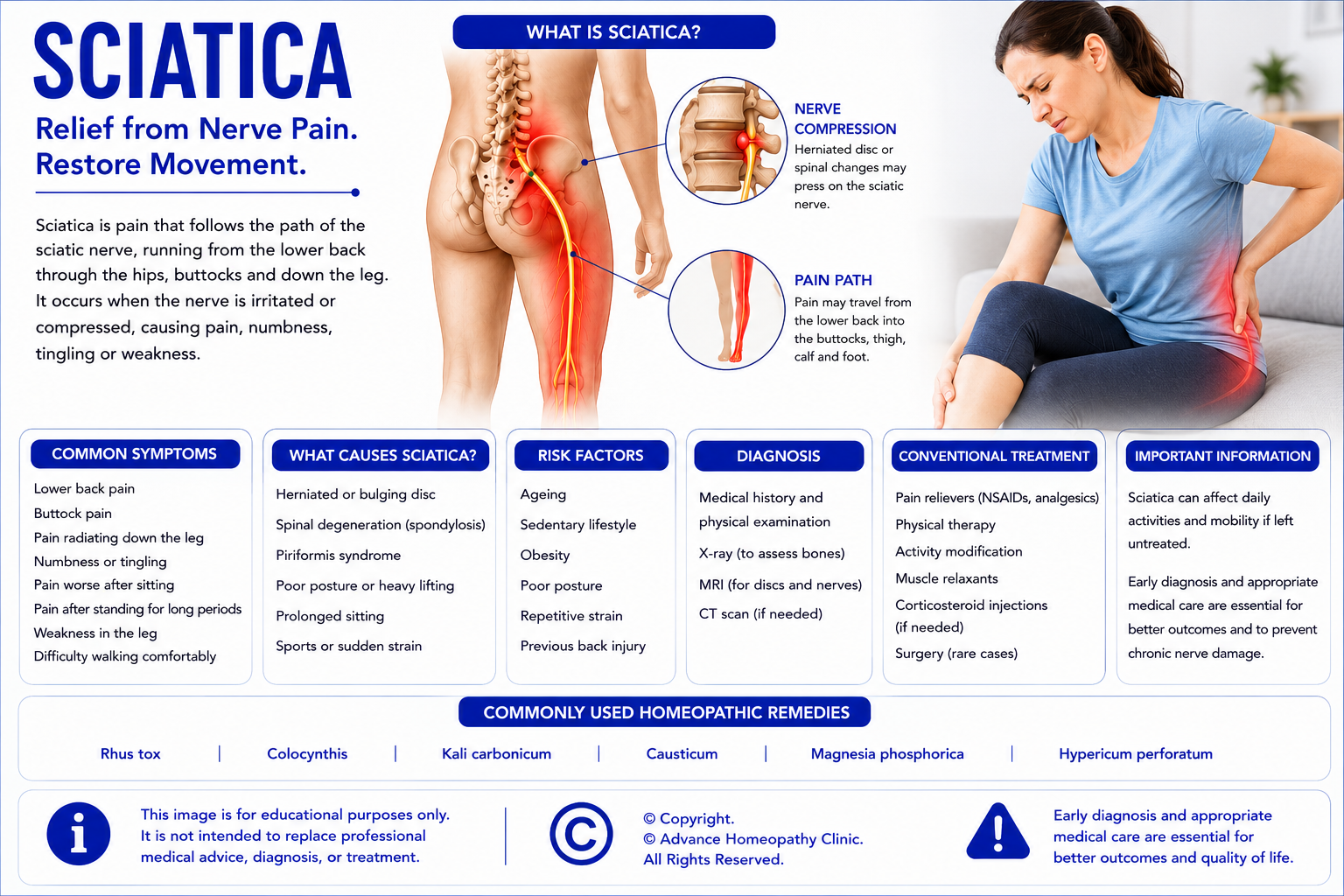
Sciatica is a condition in which pain radiates along the path of the sciatic nerve, the largest nerve in the body. The sciatic nerve originates from the lower spine and travels through the buttocks, down the back of each leg, and into the feet. Sciatica is not a disease itself but a symptom of an underlying condition that irritates or compresses one or more of the spinal nerve roots.
The most common causes include a herniated (slipped) lumbar disc, lumbar spondylosis, spinal stenosis, or other conditions that place pressure on the sciatic nerve. Depending on the severity of nerve compression, symptoms may range from mild discomfort to severe pain associated with numbness, tingling, muscle weakness, or difficulty walking. Early diagnosis is important to identify the underlying cause and guide appropriate treatment.
Common Signs & Symptoms of Sciatica
Patients with sciatica may experience one or more of the following symptoms:
- Lower back pain radiating into the buttock and leg
- Sharp, burning, or shooting pain along the course of the sciatic nerve
- Pain affecting one leg more than the other
- Numbness or tingling in the leg or foot
- Muscle weakness in the affected leg
- Pain aggravated by prolonged sitting, coughing, or sneezing
- Difficulty standing, walking, or climbing stairs
- Reduced mobility due to pain
Risk Factors for Sciatica
Several factors may increase the likelihood of developing sciatica, including:
- Herniated (slipped) lumbar disc
- Lumbar spondylosis
- Lumbar spinal stenosis
- Degenerative disc disease
- Previous spinal injury
- Repetitive lifting or bending
- Obesity
- Sedentary lifestyle
- Diabetes-related nerve damage
- Occupations involving prolonged sitting or heavy manual work
Conventional Management of Sciatica
Treatment depends on the underlying cause, severity of symptoms, and neurological findings. Most patients improve with conservative management, while some require specialist intervention.
Conventional management may include:
- Activity modification
- Physiotherapy and supervised exercise programmes
- Pain-relieving medications
- Non-steroidal anti-inflammatory drugs (NSAIDs)
- Muscle relaxants when indicated
- Neuropathic pain medications for selected patients
- Epidural steroid injections in appropriate cases
- Surgical decompression when significant nerve compression persists despite conservative treatment or when progressive neurological deficits develop
Homeopathic Medicines Commonly Considered for Sciatica
Some homeopathic medicines that may be considered by experienced homeopathic physicians for sciatica include:
- Colocynthis
- Gnaphalium Polycephalum
- Magnesia Phosphorica
- Hypericum Perforatum
- Rhus Toxicodendron
- Bryonia Alba
- Tellurium
- Kali Iodatum
Homeopathic medicines should only be prescribed by a qualified homeopathic physician after an individualized clinical evaluation.
Important: Complementary Care for Sciatica
Sciatica should always be evaluated and managed by a qualified orthopaedic surgeon, spine specialist, neurologist, rehabilitation physician, or appropriately trained healthcare professional to confirm the diagnosis, identify the underlying cause of nerve root compression, assess the severity of neurological involvement, evaluate functional impairment, and develop an individualized management plan. Comprehensive evaluation commonly includes detailed clinical history, musculoskeletal and neurological examination, assessment of lower limb muscle strength, reflexes, sensation, gait, straight leg raise and other nerve tension tests, Magnetic Resonance Imaging (MRI) of the lumbar spine, Digital X-rays where appropriate, Computed Tomography (CT) when clinically indicated, and Nerve Conduction Studies (NCS) or Electromyography (EMG) in selected patients with persistent or complex neurological symptoms. Additional investigations may be required depending on the patient's clinical presentation. Accurate diagnosis is essential because leg pain may also result from lumbar spinal stenosis, peripheral neuropathy, hip disorders, sacroiliac joint dysfunction, peripheral vascular disease, piriformis syndrome, spinal tumours, infections, or other neurological and musculoskeletal conditions, each requiring different management.
Patients with stable Sciatica may be considered for individualized homeopathic treatment as a complementary approach following appropriate orthopedic and neurological evaluation. Homeopathy may be integrated alongside physiotherapy, structured rehabilitation programmes, supervised stretching and strengthening exercises, core stabilization training, posture correction, ergonomic modification, weight management where appropriate, healthy lifestyle modification, prescribed medications, and regular follow-up with their orthopaedic surgeon or spine specialist. Individualized homeopathic prescribing is based on the patient's complete clinical presentation, constitutional characteristics, pain distribution, neurological findings, associated medical conditions, medical history, occupational demands, lifestyle factors, emotional well-being, and overall health rather than the diagnosis alone.
Patients should continue all recommended conventional treatment, including physiotherapy, structured rehabilitation, activity modification, non-steroidal anti-inflammatory drugs (NSAIDs) where appropriate, neuropathic pain medications when indicated, epidural steroid injections in carefully selected patients, multidisciplinary pain management, and lumbar decompression surgery or other spinal procedures when significant nerve root compression, persistent neurological deficit, or disabling symptoms have been identified. Homeopathy should not be regarded as a substitute for evidence-based orthopedic or neurological care, particularly in patients with large lumbar disc herniation, progressive motor weakness, severe spinal stenosis, Cauda Equina Syndrome, spinal tumours, spinal infections, or other structural disorders requiring specialist intervention.
Patients with persistent or worsening leg pain despite appropriate treatment, progressive numbness, increasing muscle weakness, recurrent falls, impaired walking ability, significant limitation of daily activities, persistent neurological deficits, or uncertainty regarding the diagnosis should continue specialist orthopedic, spine, or neurological care and regular monitoring. Homeopathy should not replace recommended imaging studies, rehabilitation programmes, spinal injections, or surgical treatment when these are advised by the treating specialist.
Patients who develop loss of bladder or bowel control, numbness around the buttocks or genital region (saddle anaesthesia), rapidly progressive weakness of one or both legs, inability to stand or walk, severe back pain following significant trauma, fever associated with severe spinal pain, or any sudden deterioration in their neurological condition should seek immediate emergency medical attention, as these symptoms may indicate Cauda Equina Syndrome, acute lumbar disc prolapse with severe nerve compression, spinal epidural abscess, spinal fracture, spinal tumour, or another serious spinal emergency requiring urgent hospital-based evaluation and treatment.
4. Frozen Shoulder (Adhesive Capsulitis)
What is Frozen Shoulder (Adhesive Capsulitis)?
Frozen shoulder, medically known as adhesive capsulitis, is a condition characterised by pain, stiffness, and progressive loss of movement of the shoulder joint. It develops when the capsule surrounding the shoulder joint becomes inflamed, thickened, and tight, restricting normal movement. The condition usually progresses gradually through different stages and may take several months or even years to resolve.
Frozen shoulder commonly affects adults between 40 and 60 years of age and is more frequently seen in people with diabetes, thyroid disorders, or following prolonged shoulder immobilisation after injury or surgery. Everyday activities such as reaching overhead, dressing, combing hair, driving, or fastening clothing may become increasingly difficult because of pain and restricted shoulder movement.

Common Signs & Symptoms of Frozen Shoulder (Adhesive Capsulitis)
Patients with frozen shoulder may experience one or more of the following symptoms:
- Shoulder pain that gradually worsens
- Progressive stiffness of the shoulder joint
- Difficulty raising the arm above shoulder level
- Pain during movement or at night
- Restricted range of shoulder movement
- Difficulty dressing, grooming, or reaching behind the back
- Difficulty sleeping on the affected side
- Reduced shoulder function during daily activities
Risk Factors for Frozen Shoulder (Adhesive Capsulitis)
Several factors may increase the likelihood of developing frozen shoulder, including:
- Diabetes mellitus
- Thyroid disorders
- Previous shoulder injury
- Shoulder surgery
- Prolonged shoulder immobilisation
- Age between 40 and 60 years
- Female gender
- Parkinson's disease
- Stroke
- Other chronic medical conditions affecting mobility
Conventional Management of Frozen Shoulder (Adhesive Capsulitis)
Treatment aims to reduce pain, restore shoulder mobility, and improve function. Recovery is often gradual and requires patience and consistent rehabilitation.
Conventional management may include:
- Physiotherapy and stretching exercises
- Home exercise programmes
- Pain-relieving medications
- Non-steroidal anti-inflammatory drugs (NSAIDs)
- Corticosteroid injections where appropriate
- Hydrodilatation in selected patients
- Manipulation under anaesthesia for resistant cases
- Arthroscopic capsular release surgery when conservative treatment has not been successful
Homeopathic Medicines Commonly Considered for Frozen Shoulder (Adhesive Capsulitis)
Some homeopathic medicines that may be considered by experienced homeopathic physicians for frozen shoulder include:
- Rhus Toxicodendron
- Bryonia Alba
- Ferrum Metallicum
- Sanguinaria Canadensis
- Ruta Graveolens
- Causticum
- Kalmia Latifolia
- Guaiacum Officinale
Homeopathic medicines should only be prescribed by a qualified homeopathic physician after an individualized clinical evaluation.
Important: Complementary Care for Frozen Shoulder (Adhesive Capsulitis)
Frozen Shoulder (Adhesive Capsulitis) should always be evaluated and managed by a qualified orthopaedic surgeon, sports medicine specialist, rehabilitation physician, or appropriately trained healthcare professional to confirm the diagnosis, determine the stage of the condition, assess the severity of pain and restriction of shoulder movement, identify underlying medical conditions, and develop an individualized rehabilitation plan. Comprehensive evaluation commonly includes detailed clinical history, physical examination assessing both active and passive range of motion, evaluation of shoulder strength, assessment for associated neck pathology, Digital X-rays to exclude bony abnormalities, ultrasound examination where appropriate, and Magnetic Resonance Imaging (MRI) when clinically indicated to exclude rotator cuff tears, shoulder arthritis, or other structural disorders. Additional investigations such as blood glucose, HbA1c, and thyroid function tests may be recommended in selected patients because Frozen Shoulder is more common in individuals with diabetes mellitus and thyroid disorders. Accurate diagnosis is essential because shoulder pain and stiffness may also result from rotator cuff disease, shoulder impingement syndrome, osteoarthritis, calcific tendinitis, cervical radiculopathy, fractures, infections, or other musculoskeletal disorders, each requiring different management.
Patients with stable Frozen Shoulder may be considered for individualized homeopathic treatment as a complementary approach following appropriate orthopedic evaluation. Homeopathy may be integrated alongside physiotherapy, supervised shoulder stretching and mobility exercises, progressive strengthening programmes, pain management, healthy lifestyle modification, management of associated medical conditions such as diabetes, prescribed medications, and regular follow-up with their orthopaedic surgeon or rehabilitation specialist. Individualized homeopathic prescribing is based on the patient's complete clinical presentation, constitutional characteristics, stage of Frozen Shoulder, associated symptoms, medical history, occupational demands, lifestyle factors, emotional well-being, and overall health rather than the diagnosis alone.
Patients should continue all recommended conventional treatment, including structured physiotherapy, home exercise programmes, pain-relieving medications, non-steroidal anti-inflammatory drugs (NSAIDs) where appropriate, intra-articular corticosteroid injections in carefully selected patients, hydrodilatation where clinically indicated, manipulation under anaesthesia or arthroscopic capsular release for persistent severe stiffness, and regular orthopedic follow-up. Homeopathy should not be regarded as a substitute for evidence-based orthopedic care, particularly in patients with marked functional limitation, persistent severe stiffness despite rehabilitation, significant structural shoulder pathology, traumatic shoulder injuries, recurrent shoulder instability, or those requiring interventional or surgical treatment.
Patients with persistent pain and stiffness despite appropriate treatment, progressive loss of shoulder function, suspected rotator cuff tear, recurrent shoulder dislocation, significant weakness, abnormal imaging findings, or uncertainty regarding the diagnosis should continue specialist orthopedic care and regular monitoring. Homeopathy should not replace recommended imaging studies, physiotherapy, rehabilitation programmes, corticosteroid injections, hydrodilatation, or surgical treatment when these are advised by the treating specialist.
Patients who develop sudden severe shoulder pain following trauma, inability to move the shoulder after injury, visible shoulder deformity, rapidly increasing swelling, fever associated with a painful shoulder, redness and warmth of the joint, numbness or weakness of the arm, loss of hand function, or any sudden deterioration in their condition should seek immediate medical attention, as these symptoms may indicate shoulder fracture, shoulder dislocation, septic arthritis, acute rotator cuff tear, nerve injury, or another serious orthopedic emergency requiring urgent hospital-based evaluation and treatment.
5. Carpal Tunnel Syndrome
What is Carpal Tunnel Syndrome?
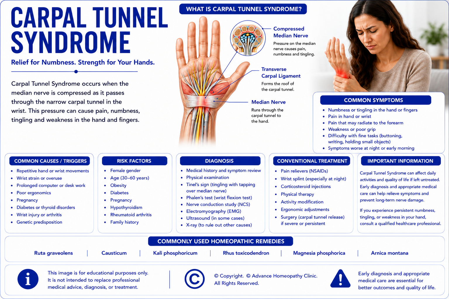
Carpal Tunnel Syndrome (CTS) is a common nerve compression disorder that occurs when the median nerve becomes compressed as it passes through the carpal tunnel—a narrow passageway in the wrist formed by bones and ligaments. The median nerve supplies sensation to the thumb, index finger, middle finger, and part of the ring finger, while also controlling several muscles responsible for thumb movement.
Compression of the median nerve may result from swelling of the surrounding tissues, repetitive hand movements, wrist injuries, inflammatory conditions, pregnancy, diabetes, thyroid disorders, or other medical conditions. Symptoms usually develop gradually and may initially occur only at night before progressing to persistent numbness, tingling, pain, weakness, or reduced hand function. Early diagnosis and appropriate treatment are important to prevent permanent nerve damage and preserve hand strength.
Common Signs & Symptoms of Carpal Tunnel Syndrome
Patients with Carpal Tunnel Syndrome may experience one or more of the following symptoms:
- Numbness or tingling of the thumb, index, middle, and ring fingers
- Burning or aching pain in the wrist or hand
- Pain radiating into the forearm
- Symptoms that are worse at night
- Weakness of hand grip
- Difficulty holding or grasping objects
- Frequent dropping of objects
- Reduced thumb strength
- Relief of symptoms after shaking the hand (flick sign)
Risk Factors for Carpal Tunnel Syndrome
Several factors may increase the likelihood of developing Carpal Tunnel Syndrome, including:
- Repetitive hand and wrist movements
- Prolonged keyboard or computer use
- Diabetes mellitus
- Hypothyroidism
- Pregnancy
- Rheumatoid arthritis
- Wrist fractures or previous wrist injuries
- Obesity
- Occupations involving repetitive gripping or vibrating tools
- Female gender
Conventional Management of Carpal Tunnel Syndrome
Treatment depends on symptom severity, duration, and the degree of nerve compression. Early treatment often prevents progression and improves long-term outcomes.
Conventional management may include:
- Activity modification
- Wrist splinting, particularly at night
- Ergonomic workplace modifications
- Physiotherapy and nerve-gliding exercises
- Pain-relieving medications
- Non-steroidal anti-inflammatory drugs (NSAIDs) where appropriate
- Corticosteroid injections for selected patients
- Nerve conduction studies (NCS) and electromyography (EMG) when indicated
- Surgical carpal tunnel release for persistent or severe nerve compression
Homeopathic Medicines Commonly Considered for Carpal Tunnel Syndrome
Some homeopathic medicines that may be considered by experienced homeopathic physicians for Carpal Tunnel Syndrome include:
- Hypericum Perforatum
- Ruta Graveolens
- Causticum
- Rhus Toxicodendron
- Arnica Montana
- Guaiacum Officinale
- Bryonia Alba
- Kalmia Latifolia
Homeopathic medicines should only be prescribed by a qualified homeopathic physician after an individualized clinical evaluation.
Important: Complementary Care for Carpal Tunnel Syndrome
Carpal Tunnel Syndrome (CTS) should always be evaluated and managed by a qualified orthopaedic surgeon, hand surgeon, neurologist, rehabilitation physician, or appropriately trained healthcare professional to confirm the diagnosis, determine the severity of median nerve compression, identify contributing medical conditions, assess functional impairment, and develop an individualized management plan. Comprehensive evaluation commonly includes detailed clinical history, physical examination of the hand and wrist, assessment of sensory function, thumb muscle strength, grip strength, provocative clinical tests where appropriate, Nerve Conduction Studies (NCS), Electromyography (EMG), ultrasound examination of the median nerve when clinically indicated, and additional imaging studies where necessary to exclude other wrist disorders. Additional investigations such as blood glucose, HbA1c, thyroid function tests, and inflammatory markers may be recommended in selected patients because Carpal Tunnel Syndrome may be associated with diabetes mellitus, hypothyroidism, rheumatoid arthritis, pregnancy, obesity, or repetitive occupational hand use. Accurate diagnosis is essential because hand numbness and weakness may also result from cervical radiculopathy, peripheral neuropathy, pronator syndrome, ulnar neuropathy, wrist arthritis, tendon disorders, or other neurological and musculoskeletal conditions, each requiring different management.
Patients with mild to moderate Carpal Tunnel Syndrome may be considered for individualized homeopathic treatment as a complementary approach following appropriate orthopedic or neurological evaluation. Homeopathy may be integrated alongside activity modification, ergonomic workplace adjustments, wrist splinting, physiotherapy, tendon and nerve gliding exercises, healthy lifestyle modification, management of underlying medical conditions, prescribed medications, and regular follow-up with their orthopaedic surgeon, neurologist, or hand specialist. Individualized homeopathic prescribing is based on the patient's complete clinical presentation, constitutional characteristics, symptom pattern, severity of nerve compression, associated medical conditions, occupational demands, lifestyle factors, emotional well-being, and overall health rather than the diagnosis alone.
Patients should continue all recommended conventional treatment, including wrist splinting, activity modification, ergonomic correction, physiotherapy, non-steroidal anti-inflammatory drugs (NSAIDs) where appropriate, corticosteroid injections in carefully selected patients, treatment of underlying medical conditions, and carpal tunnel release surgery when persistent symptoms, significant nerve compression, progressive weakness, or muscle wasting has been identified. Homeopathy should not be regarded as a substitute for evidence-based orthopedic or neurological care, particularly in patients with severe median nerve compression, persistent numbness, progressive weakness, thenar muscle wasting, significant functional impairment, or those requiring surgical decompression.
Patients with persistent symptoms despite appropriate treatment, worsening numbness, increasing weakness of thumb movements, reduced grip strength, muscle wasting at the base of the thumb, abnormal NCS or EMG findings, recurrent symptoms following treatment, or uncertainty regarding the diagnosis should continue specialist orthopedic, hand surgery, or neurological care and regular monitoring. Homeopathy should not replace recommended NCS, EMG, rehabilitation programmes, corticosteroid injections, or surgical decompression when these are advised by the treating specialist.
Patients who develop sudden inability to move the thumb or fingers, rapidly progressive hand weakness, severe hand trauma, marked swelling associated with severe pain, signs of infection such as fever with redness and warmth of the hand, sudden loss of sensation affecting the entire hand, or any sudden deterioration in hand function should seek immediate medical attention, as these symptoms may indicate acute nerve injury, compartment syndrome, severe traumatic injury, septic arthritis, flexor tendon injury, or another serious orthopedic or neurological emergency requiring urgent hospital-based evaluation and treatment.
6. De Quervain's Tenosynovitis
What is De Quervain's Tenosynovitis?
De Quervain's Tenosynovitis is a painful condition affecting the tendons that control movement of the thumb as they pass through a narrow tunnel on the thumb side of the wrist. Repetitive movements, overuse, or irritation of these tendons may cause inflammation and thickening of the tendon sheath, making thumb and wrist movements painful.
The condition commonly affects individuals who frequently perform repetitive gripping, lifting, twisting, or pinching activities. It is also frequently seen in new parents due to repeated lifting of infants, as well as in people whose occupations or sports require repetitive thumb and wrist movements. Pain may gradually worsen and interfere with activities such as opening jars, lifting objects, writing, typing, or using a mobile phone.

Common Signs & Symptoms of De Quervain's Tenosynovitis
Patients with De Quervain's Tenosynovitis may experience one or more of the following symptoms:
- Pain on the thumb side of the wrist
- Pain aggravated by thumb movement or gripping
- Swelling near the base of the thumb
- Difficulty gripping or pinching objects
- Pain while lifting or carrying objects
- Clicking or snapping sensation during thumb movement
- Reduced thumb strength
- Wrist stiffness
Risk Factors for De Quervain's Tenosynovitis
Several factors may increase the likelihood of developing De Quervain's Tenosynovitis, including:
- Repetitive thumb movements
- Frequent lifting of infants or young children
- Repetitive gripping or twisting activities
- Gardening or racquet sports
- Occupational overuse
- Pregnancy and the postpartum period
- Inflammatory arthritis
- Previous wrist injury
- Female gender
- Age between 30 and 50 years
Conventional Management of De Quervain's Tenosynovitis
Treatment focuses on reducing tendon inflammation, relieving pain, and restoring normal thumb function.
Conventional management may include:
- Activity modification
- Thumb spica splinting
- Physiotherapy
- Tendon-gliding exercises
- Pain-relieving medications
- Non-steroidal anti-inflammatory drugs (NSAIDs)
- Corticosteroid injections where indicated
- Ultrasound-guided injections for selected patients
- Surgical release of the tendon sheath for persistent cases
Homeopathic Medicines Commonly Considered for De Quervain's Tenosynovitis
Some homeopathic medicines that may be considered by experienced homeopathic physicians for De Quervain's Tenosynovitis include:
- Ruta Graveolens
- Rhus Toxicodendron
- Bryonia Alba
- Arnica Montana
- Hypericum Perforatum
- Causticum
- Guaiacum Officinale
- Calcarea Fluorica
Homeopathic medicines should only be prescribed by a qualified homeopathic physician after an individualized clinical evaluation.
Important: Complementary Care for De Quervain's Tenosynovitis
De Quervain's Tenosynovitis should always be evaluated and managed by a qualified orthopaedic surgeon, hand surgeon, sports medicine specialist, rehabilitation physician, or appropriately trained healthcare professional to confirm the diagnosis, determine the severity of tendon involvement, identify contributing occupational or repetitive activities, assess functional impairment, and develop an individualized management plan. Comprehensive evaluation commonly includes detailed clinical history, physical examination of the thumb and wrist, assessment of grip strength and thumb movement, evaluation of tenderness over the first dorsal compartment, provocative clinical tests such as Finkelstein's test where appropriate, ultrasound examination when clinically indicated to assess tendon sheath inflammation, and additional imaging studies to exclude fractures, thumb carpometacarpal arthritis, or other wrist disorders when necessary. Accurate diagnosis is essential because pain over the thumb side of the wrist may also result from thumb osteoarthritis, wrist sprains, tendon injuries, intersection syndrome, scaphoid fractures, radial nerve disorders, or other musculoskeletal conditions, each requiring different management.
Patients with mild to moderate De Quervain's Tenosynovitis may be considered for individualized homeopathic treatment as a complementary approach following appropriate orthopedic evaluation. Homeopathy may be integrated alongside activity modification, avoidance of repetitive thumb movements, ergonomic workplace adjustments, thumb spica splinting, physiotherapy, supervised tendon gliding and strengthening exercises, healthy lifestyle modification, prescribed medications, and regular follow-up with their orthopaedic surgeon, hand specialist, or rehabilitation physician. Individualized homeopathic prescribing is based on the patient's complete clinical presentation, constitutional characteristics, symptom pattern, severity of tendon involvement, occupational demands, associated medical conditions, lifestyle factors, emotional well-being, and overall health rather than the diagnosis alone.
Patients should continue all recommended conventional treatment, including thumb spica splinting, activity modification, ergonomic correction, physiotherapy, non-steroidal anti-inflammatory drugs (NSAIDs) where appropriate, corticosteroid injections in carefully selected patients, structured rehabilitation programmes, and surgical release of the first dorsal compartment when persistent pain, significant functional limitation, or failure of conservative treatment has been identified. Homeopathy should not be regarded as a substitute for evidence-based orthopedic care, particularly in patients with persistent severe pain, marked loss of thumb function, significant tendon thickening, recurrent symptoms despite conservative treatment, or those requiring corticosteroid injection or surgical intervention.
Patients with persistent pain despite appropriate treatment, progressive loss of thumb movement, increasing weakness of grip, recurrent symptoms affecting work or daily activities, abnormal imaging findings, or uncertainty regarding the diagnosis should continue specialist orthopedic or hand surgery care and regular monitoring. Homeopathy should not replace recommended imaging studies, splinting, rehabilitation programmes, corticosteroid injections, or surgical treatment when these are advised by the treating specialist.
Patients who develop sudden inability to move the thumb following injury, severe wrist trauma, visible deformity, rapidly increasing swelling, severe redness and warmth associated with fever, loss of sensation affecting the hand or thumb, or any sudden deterioration in hand function should seek immediate medical attention, as these symptoms may indicate tendon rupture, fracture, septic tenosynovitis, acute nerve injury, compartment syndrome, or another serious orthopedic emergency requiring urgent hospital-based evaluation and treatment.
7. Plantar Fasciitis
What is Plantar Fasciitis?
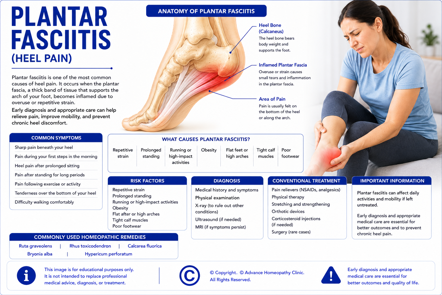
Plantar fasciitis is one of the most common causes of heel pain. It occurs when the plantar fascia, a strong band of connective tissue that supports the arch of the foot and connects the heel bone to the toes, becomes irritated or undergoes repetitive microscopic injury. Although inflammation may be present during the early stages, many chronic cases involve degeneration of the plantar fascia rather than ongoing inflammation.
The condition commonly develops due to repetitive strain placed on the plantar fascia from prolonged standing, excessive walking or running, obesity, poor foot mechanics, or inappropriate footwear. Patients typically experience sharp pain beneath the heel during the first few steps after getting out of bed in the morning or after prolonged periods of rest. Early diagnosis and appropriate treatment can help relieve pain, restore mobility, and prevent long-term disability.
Common Signs & Symptoms of Plantar Fasciitis
Patients with plantar fasciitis may experience one or more of the following symptoms:
- Sharp pain beneath the heel
- Heel pain during the first steps in the morning
- Pain after prolonged sitting or rest
- Pain that improves after walking for a short time but returns with prolonged activity
- Heel pain after prolonged standing
- Pain after walking or running
- Tenderness over the bottom of the heel
- Difficulty walking comfortably
Risk Factors for Plantar Fasciitis
Several factors may increase the likelihood of developing plantar fasciitis, including:
- Prolonged standing
- Running and other high-impact sports
- Obesity
- Flat feet (pes planus)
- High foot arches (pes cavus)
- Tight calf muscles or Achilles tendon
- Poorly supportive footwear
- Occupations requiring prolonged standing or walking
- Increasing age
Conventional Management of Plantar Fasciitis
Treatment focuses on reducing pain, allowing the plantar fascia to heal, and correcting contributing biomechanical factors.
Conventional management may include:
- Activity modification
- Stretching exercises for the plantar fascia and calf muscles
- Physiotherapy
- Supportive footwear
- Custom orthotic insoles where indicated
- Ice application
- Pain-relieving medications
- Non-steroidal anti-inflammatory drugs (NSAIDs)
- Extracorporeal shockwave therapy (ESWT) for selected patients
- Corticosteroid injections in carefully selected cases
- Surgical release for persistent symptoms that fail conservative treatment
Homeopathic Medicines Commonly Considered for Plantar Fasciitis
Some homeopathic medicines that may be considered by experienced homeopathic physicians for plantar fasciitis include:
- Ruta Graveolens
- Rhus Toxicodendron
- Bryonia Alba
- Calcarea Fluorica
- Hypericum Perforatum
- Arnica Montana
- Guaiacum Officinale
- Hekla Lava
Homeopathic medicines should only be prescribed by a qualified homeopathic physician after an individualized clinical evaluation.
Important: Complementary Care for Plantar Fasciitis
Plantar Fasciitis should always be evaluated and managed by a qualified orthopaedic surgeon, foot and ankle specialist, sports medicine specialist, rehabilitation physician, or appropriately trained healthcare professional to confirm the diagnosis, determine the severity of plantar fascia involvement, identify contributing biomechanical factors, assess functional impairment, and develop an individualized management plan. Comprehensive evaluation commonly includes detailed clinical history, physical examination of the foot and ankle, assessment of gait, foot posture, ankle flexibility, calf muscle tightness, and tenderness at the plantar fascia insertion, together with Digital X-rays to exclude fractures or significant bony abnormalities, ultrasound examination where appropriate to assess plantar fascia thickening, and Magnetic Resonance Imaging (MRI) when clinically indicated to exclude alternative causes of persistent heel pain. Additional investigations may be recommended depending on the patient's clinical presentation. Accurate diagnosis is essential because heel pain may also result from calcaneal stress fractures, nerve entrapment syndromes, inflammatory arthritis, Achilles tendon disorders, plantar fascia rupture, infections, tumours, or other musculoskeletal conditions, each requiring different management.
Patients with stable Plantar Fasciitis may be considered for individualized homeopathic treatment as a complementary approach following appropriate orthopedic evaluation. Homeopathy may be integrated alongside activity modification, supervised stretching exercises for the plantar fascia and calf muscles, strengthening exercises for the foot and ankle, physiotherapy, weight management where appropriate, supportive footwear, orthotic devices when indicated, healthy lifestyle modification, prescribed medications, and regular follow-up with their orthopaedic surgeon, sports medicine specialist, or rehabilitation physician. Individualized homeopathic prescribing is based on the patient's complete clinical presentation, constitutional characteristics, pain pattern, functional limitations, occupational demands, associated medical conditions, lifestyle factors, emotional well-being, and overall health rather than the diagnosis alone.
Patients should continue all recommended conventional treatment, including structured physiotherapy, plantar fascia and calf stretching programmes, strengthening exercises, activity modification, appropriate footwear, custom orthoses where indicated, night splints in selected patients, non-steroidal anti-inflammatory drugs (NSAIDs) where appropriate, extracorporeal shockwave therapy (ESWT) for carefully selected chronic cases, corticosteroid injections or other image-guided interventions in selected patients, and surgical treatment only when persistent disabling symptoms fail to respond to prolonged conservative management. Homeopathy should not be regarded as a substitute for evidence-based orthopedic care, particularly in patients with persistent severe pain, plantar fascia rupture, stress fractures, inflammatory arthritis, significant structural foot abnormalities, or those requiring interventional or surgical treatment.
Patients with persistent heel pain despite appropriate treatment, progressive limitation of walking, recurrent symptoms affecting work or sporting activities, abnormal imaging findings, suspected inflammatory or systemic disease, or uncertainty regarding the diagnosis should continue specialist orthopedic or foot and ankle care and regular monitoring. Homeopathy should not replace recommended imaging studies, physiotherapy, orthotic management, extracorporeal shockwave therapy, image-guided interventions, or surgical treatment when these are advised by the treating specialist.
Patients who develop sudden severe heel pain associated with a popping sensation, inability to bear weight following injury, marked swelling, visible deformity, fever associated with redness and warmth of the foot, rapidly progressive neurological symptoms affecting the foot, or any sudden deterioration in their ability to walk should seek immediate medical attention, as these symptoms may indicate plantar fascia rupture, calcaneal fracture, septic arthritis, osteomyelitis, acute tendon injury, compartment syndrome, or another serious orthopedic emergency requiring urgent hospital-based evaluation and treatment.
8. Calcaneal Spur (Heel Spur)
What is Calcaneal Spur (Heel Spur)?
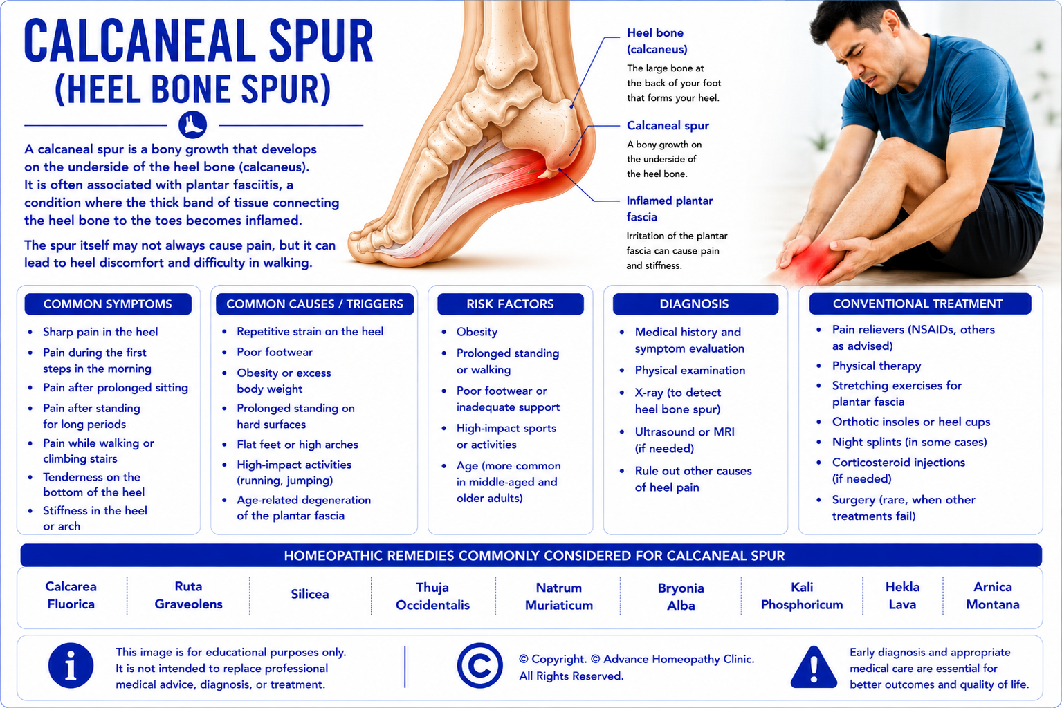
A calcaneal spur, commonly known as a heel spur, is a small bony outgrowth that develops on the underside or back of the heel bone (calcaneus). Heel spurs usually develop gradually over time due to repetitive traction where the plantar fascia or Achilles tendon attaches to the heel bone. Although heel spurs are frequently associated with plantar fasciitis, many people have heel spurs without experiencing any symptoms.
When symptoms occur, pain is often caused by the associated soft tissue irritation rather than the spur itself. Patients may experience discomfort while standing, walking, or placing weight on the heel, particularly after periods of rest. Proper medical evaluation is important because heel pain may arise from several different orthopedic conditions that require different management strategies.
Common Signs & Symptoms of Calcaneal Spur (Heel Spur)
Patients with a calcaneal spur may experience one or more of the following symptoms:
- Pain beneath or behind the heel
- Pain during the first steps after resting
- Heel pain while standing or walking
- Tenderness over the heel bone
- Pain after prolonged standing
- Discomfort during weight-bearing activities
- Difficulty walking long distances
- Localised heel tenderness
Risk Factors for Calcaneal Spur (Heel Spur)
Several factors may increase the likelihood of developing a calcaneal spur, including:
- Long-standing plantar fasciitis
- Repetitive stress on the heel
- Obesity
- Flat feet
- High foot arches
- Tight calf muscles
- Poor footwear
- Occupations requiring prolonged standing
- Increasing age
Conventional Management of Calcaneal Spur (Heel Spur)
Treatment aims to reduce pain, minimise stress on the heel, and improve walking ability.
Conventional management may include:
- Activity modification
- Stretching exercises
- Physiotherapy
- Supportive footwear
- Orthotic insoles
- Heel cushions
- Pain-relieving medications
- Non-steroidal anti-inflammatory drugs (NSAIDs)
- Extracorporeal shockwave therapy (ESWT)
- Corticosteroid injections for selected patients
- Surgical removal in carefully selected persistent cases
Homeopathic Medicines Commonly Considered for Calcaneal Spur (Heel Spur)
Some homeopathic medicines that may be considered by experienced homeopathic physicians for calcaneal spur include:
- Hekla Lava
- Ruta Graveolens
- Calcarea Fluorica
- Rhus Toxicodendron
- Bryonia Alba
- Hypericum Perforatum
- Arnica Montana
- Guaiacum Officinale
Homeopathic medicines should only be prescribed by a qualified homeopathic physician after an individualized clinical evaluation.
Important: Complementary Care for Calcaneal Spur (Heel Spur)
Calcaneal Spur (Heel Spur) should always be evaluated and managed by a qualified orthopaedic surgeon, foot and ankle specialist, sports medicine specialist, rehabilitation physician, or appropriately trained healthcare professional to confirm the diagnosis, determine whether the heel spur is responsible for the patient's symptoms, identify associated plantar fasciitis or other foot disorders, assess functional impairment, and develop an individualized management plan. Comprehensive evaluation commonly includes detailed clinical history, physical examination of the foot and ankle, assessment of gait, foot biomechanics, ankle flexibility, calf muscle tightness, tenderness at the plantar fascia insertion, Digital X-rays to identify calcaneal spurs and exclude fractures, ultrasound examination where appropriate to evaluate plantar fascia pathology, and Magnetic Resonance Imaging (MRI) when clinically indicated to investigate persistent or atypical heel pain. Additional investigations may be recommended depending on the patient's clinical presentation. Accurate diagnosis is essential because heel pain may also result from plantar fasciitis, calcaneal stress fractures, Achilles tendon disorders, nerve entrapment syndromes, inflammatory arthritis, infections, tumours, or other musculoskeletal conditions, each requiring different management.
Patients with stable Calcaneal Spur may be considered for individualized homeopathic treatment as a complementary approach following appropriate orthopedic evaluation. Homeopathy may be integrated alongside activity modification, physiotherapy, supervised stretching of the plantar fascia and calf muscles, strengthening exercises, weight management where appropriate, supportive footwear, custom orthotic devices when indicated, healthy lifestyle modification, prescribed medications, and regular follow-up with their orthopaedic surgeon or rehabilitation specialist. Individualized homeopathic prescribing is based on the patient's complete clinical presentation, constitutional characteristics, pain pattern, biomechanical abnormalities, functional limitations, associated medical conditions, occupational demands, lifestyle factors, emotional well-being, and overall health rather than the diagnosis alone.
Patients should continue all recommended conventional treatment, including structured physiotherapy, plantar fascia rehabilitation programmes, stretching and strengthening exercises, footwear modification, orthotic support where indicated, non-steroidal anti-inflammatory drugs (NSAIDs) where appropriate, extracorporeal shockwave therapy (ESWT) for carefully selected chronic cases, image-guided corticosteroid injections in selected patients, and surgical excision of the heel spur with plantar fascia release only in carefully selected patients whose symptoms remain disabling despite prolonged conservative treatment. Homeopathy should not be regarded as a substitute for evidence-based orthopedic care, particularly in patients with persistent severe heel pain, plantar fascia rupture, calcaneal stress fractures, inflammatory arthritis, significant structural foot abnormalities, or those requiring interventional or surgical treatment.
Patients with persistent heel pain despite appropriate treatment, progressive limitation of walking, recurrent symptoms affecting work or sporting activities, abnormal imaging findings, suspected inflammatory or systemic disease, or uncertainty regarding the diagnosis should continue specialist orthopedic or foot and ankle care and regular monitoring. Homeopathy should not replace recommended imaging studies, physiotherapy, orthotic management, extracorporeal shockwave therapy, image-guided interventions, or surgical treatment when these are advised by the treating specialist.
Patients who develop sudden severe heel pain following trauma, inability to bear weight, marked swelling, visible deformity, fever associated with redness and warmth of the foot, rapidly progressive numbness or weakness affecting the foot, or any sudden deterioration in their ability to walk should seek immediate medical attention, as these symptoms may indicate calcaneal fracture, plantar fascia rupture, septic arthritis, osteomyelitis, acute tendon injury, compartment syndrome, or another serious orthopedic emergency requiring urgent hospital-based evaluation and treatment.
9. Tennis Elbow (Lateral Epicondylitis)
What is Tennis Elbow (Lateral Epicondylitis)?
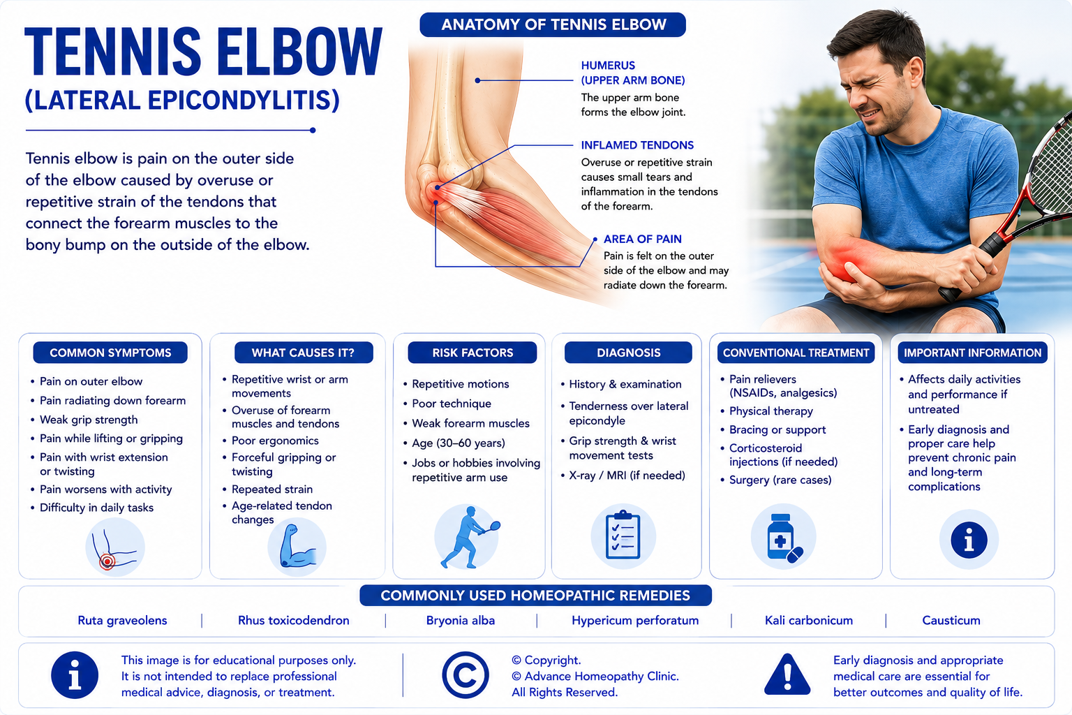
Tennis elbow, medically known as lateral epicondylitis, is a common overuse condition affecting the tendons that attach the forearm extensor muscles to the outer part of the elbow (lateral epicondyle). Despite its name, the condition affects many people who have never played tennis. It commonly develops because of repetitive gripping, lifting, twisting, typing, manual work, gardening, or sporting activities that repeatedly strain the forearm muscles and tendons.
Current research suggests that chronic tennis elbow is primarily a degenerative tendinopathy rather than a purely inflammatory condition. Repetitive microtrauma may lead to structural changes within the tendon, causing persistent pain and reduced function. Early diagnosis and appropriate treatment can help relieve pain, restore strength, and prevent long-term tendon damage.
Common Signs & Symptoms of Tennis Elbow (Lateral Epicondylitis)
Patients with tennis elbow may experience one or more of the following symptoms:
- Pain over the outer part of the elbow
- Pain during gripping activities
- Reduced grip strength
- Pain while lifting or carrying objects
- Pain during twisting movements, such as opening jars or turning door handles
- Tenderness over the lateral epicondyle
- Pain radiating into the forearm
- Difficulty performing repetitive hand activities
Risk Factors for Tennis Elbow (Lateral Epicondylitis)
Several factors may increase the likelihood of developing tennis elbow, including:
- Repetitive gripping
- Repetitive lifting
- Racquet sports
- Manual labour
- Computer and office work
- Gardening
- Plumbing, painting, carpentry, or mechanical work
- Occupational overuse
- Age between 35 and 55 years
Conventional Management of Tennis Elbow (Lateral Epicondylitis)
Treatment aims to reduce pain, promote tendon healing, restore strength, and improve arm function.
Conventional management may include:
- Activity modification
- Physiotherapy
- Eccentric strengthening exercises
- Stretching programmes
- Counterforce braces
- Pain-relieving medications
- Non-steroidal anti-inflammatory drugs (NSAIDs)
- Corticosteroid injections in selected cases
- Platelet-rich plasma (PRP) therapy for selected patients
- Extracorporeal shockwave therapy (ESWT)
- Surgical treatment for persistent cases that fail conservative management
Homeopathic Medicines Commonly Considered for Tennis Elbow (Lateral Epicondylitis)
Some homeopathic medicines that may be considered by experienced homeopathic physicians for tennis elbow include:
- Ruta Graveolens
- Rhus Toxicodendron
- Bryonia Alba
- Arnica Montana
- Hypericum Perforatum
- Guaiacum Officinale
- Causticum
- Calcarea Fluorica
Homeopathic medicines should only be prescribed by a qualified homeopathic physician after an individualized clinical evaluation.
Important: Complementary Care for Tennis Elbow (Lateral Epicondylitis)
Tennis Elbow (Lateral Epicondylitis) should always be evaluated and managed by a qualified orthopaedic surgeon, sports medicine specialist, rehabilitation physician, or appropriately trained healthcare professional to confirm the diagnosis, determine the severity of tendon involvement, identify contributing occupational or sporting activities, assess functional impairment, and develop an individualized management plan. Comprehensive evaluation commonly includes detailed clinical history, physical examination of the elbow, wrist, and forearm, assessment of grip strength, evaluation of pain during resisted wrist extension and forearm movements, examination of elbow range of motion, ultrasound examination where clinically indicated to evaluate tendon pathology, and Magnetic Resonance Imaging (MRI) in selected patients with persistent symptoms or when alternative diagnoses are suspected. Additional investigations may be recommended depending on the patient's clinical presentation. Accurate diagnosis is essential because lateral elbow pain may also result from radial tunnel syndrome, cervical radiculopathy, elbow osteoarthritis, ligament injuries, elbow instability, fractures, inflammatory arthritis, tendon tears, or other musculoskeletal and neurological conditions, each requiring different management.
Patients with mild to moderate Tennis Elbow may be considered for individualized homeopathic treatment as a complementary approach following appropriate orthopedic evaluation. Homeopathy may be integrated alongside activity modification, ergonomic workplace adjustments, supervised physiotherapy, progressive tendon loading and strengthening exercises, stretching programmes, healthy lifestyle modification, bracing where appropriate, prescribed medications, and regular follow-up with their orthopaedic surgeon, sports medicine specialist, or rehabilitation physician. Individualized homeopathic prescribing is based on the patient's complete clinical presentation, constitutional characteristics, pain pattern, severity of tendon involvement, occupational and sporting demands, associated medical conditions, lifestyle factors, emotional well-being, and overall health rather than the diagnosis alone.
Patients should continue all recommended conventional treatment, including activity modification, structured physiotherapy, eccentric and progressive resistance exercise programmes, ergonomic correction, counterforce bracing or wrist splinting where appropriate, non-steroidal anti-inflammatory drugs (NSAIDs) for short-term pain relief when clinically indicated, image-guided interventions in carefully selected patients, extracorporeal shockwave therapy (ESWT) where appropriate, and surgical treatment only for persistent disabling symptoms that fail to respond to prolonged comprehensive conservative management. Homeopathy should not be regarded as a substitute for evidence-based orthopedic care, particularly in patients with persistent severe pain, significant tendon tears, marked functional impairment, elbow instability, progressive weakness, or those requiring interventional or surgical treatment.
Patients with persistent elbow pain despite appropriate treatment, progressive reduction in grip strength, inability to perform occupational or sporting activities, recurrent symptoms despite rehabilitation, abnormal imaging findings, suspected tendon rupture, or uncertainty regarding the diagnosis should continue specialist orthopedic or sports medicine care and regular monitoring. Homeopathy should not replace recommended imaging studies, physiotherapy, rehabilitation programmes, image-guided interventions, or surgical treatment when these are advised by the treating specialist.
Patients who develop sudden severe elbow pain following trauma, inability to move the elbow, visible deformity, rapidly increasing swelling, fever associated with redness and warmth of the elbow, sudden loss of hand or wrist function, progressive numbness or weakness affecting the arm, or any sudden deterioration in upper limb function should seek immediate medical attention, as these symptoms may indicate elbow fracture, tendon rupture, septic arthritis, acute nerve injury, compartment syndrome, or another serious orthopedic emergency requiring urgent hospital-based evaluation and treatment.
A Note About Homeopathic Medicines
Homeopathy is an individualized system of medicine. Unlike conventional medicine, where patients with the same diagnosis often receive similar treatments, homeopathic medicine selection is based on the individual's overall symptom pattern rather than the diagnosis alone.
Two patients with the same orthopedic condition—for example, cervical spondylosis, plantar fasciitis, or frozen shoulder—may require different homeopathic medicines depending on factors such as:
- The nature, location, and character of the pain
- Factors that aggravate or relieve symptoms
- Associated musculoskeletal and general symptoms
- Medical history
- Individual constitutional characteristics
- Emotional and psychological factors where clinically relevant
- Overall health status
For this reason, homeopathic medicines should always be prescribed by a qualified homeopathic physician following a comprehensive clinical assessment. Self-medication may delay appropriate diagnosis and treatment, particularly in conditions requiring urgent orthopedic or neurological evaluation.
Homeopathy should be regarded as a complementary approach and should not replace conventional investigations, prescription medications, physiotherapy, rehabilitation programmes, or surgical treatment whenever these are medically indicated.
Our Homeopathic Approach
At Advance Homeopathy Clinic Dubai, every patient undergoes a detailed and individualized consultation before any treatment plan is recommended. We recognise that musculoskeletal pain often affects far more than a single joint or body part—it can influence mobility, sleep, work performance, physical activity, emotional well-being, and overall quality of life.
Our consultation includes a comprehensive evaluation of:
- The patient's presenting orthopedic condition
- Evolution and progression of symptoms
- Pain characteristics and individual symptom patterns
- Aggravating and relieving factors
- Previous injuries, surgeries, and treatments
- Imaging reports and laboratory investigations where available
- Medical and family history
- Lifestyle and occupational factors
- Overall physical and constitutional health
Where appropriate, we work alongside orthopedic surgeons, spine specialists, sports medicine physicians, physiotherapists, neurologists, rheumatologists, and primary care physicians to ensure that patients continue receiving appropriate evidence-based medical care.
Our aim is to provide individualized homeopathic treatment as part of a comprehensive and patient-centred approach to musculoskeletal health.
Why Choose Advance Homeopathy Clinic Dubai?
Choosing the right healthcare provider is an important decision. At Advance Homeopathy Clinic Dubai, we are committed to delivering ethical, patient-centred, and individualized homeopathic care while encouraging appropriate conventional medical evaluation whenever required.
Our practice is built on the following principles:
- Individualized treatment based on each patient's complete clinical presentation
- Comprehensive consultations rather than diagnosis-based prescribing
- Experienced DHA-licensed homeopathic physicians
- Care for both acute and chronic musculoskeletal conditions
- Evidence-informed clinical decision-making
- Collaboration with conventional healthcare providers whenever appropriate
- In-clinic and online consultation facilities
- Patients from numerous nationalities and countries have consulted our clinic
- A strong commitment to professional ethics, patient education, and informed decision-making
Every patient is unique, and treatment recommendations are tailored to the individual's clinical needs, medical history, and treatment goals.
Frequently Asked Questions (FAQs)
Can homeopathy treat orthopedic disorders?
Homeopathy is commonly used as a complementary approach by patients with orthopedic conditions. Treatment is individualized and should be integrated with appropriate conventional medical care. The expected benefits and suitability of homeopathy vary depending on the condition, severity, and individual patient.
Can homeopathy replace surgery?
No. Homeopathy should not be considered a substitute for surgery when surgical treatment is medically indicated. Patients should always follow the advice of their orthopedic surgeon or treating specialist.
Can I continue my conventional medications while taking homeopathic treatment?
Yes. Patients should continue all prescribed medications unless advised otherwise by their treating physician. Homeopathic treatment is commonly used alongside conventional medical care.
How long does treatment usually take?
The duration of treatment varies depending on the diagnosis, severity of the condition, duration of symptoms, underlying pathology, previous treatments, and individual response. Your homeopathic physician will discuss realistic expectations after your clinical evaluation.
Do I need investigations before starting treatment?
Existing investigations such as X-rays, MRI scans, CT scans, ultrasound reports, blood tests, or nerve conduction studies are helpful when available. If additional investigations are clinically indicated, patients may be advised to undergo appropriate evaluation before or during treatment.
Is homeopathy suitable for sports injuries?
Some patients seek homeopathic treatment as a complementary approach for selected sports-related musculoskeletal conditions. However, acute injuries, fractures, tendon ruptures, ligament tears, joint dislocations, or severe trauma require prompt orthopedic assessment and evidence-based medical management.
When should I seek urgent medical attention?
Immediate medical assessment is recommended if you experience:
- Sudden inability to move a limb
- Severe pain following significant trauma
- Suspected fracture or joint dislocation
- Progressive muscle weakness
- Loss of sensation
- Loss of bladder or bowel control
- Numbness around the groin or saddle area
- Fever associated with severe joint pain or swelling
- Rapidly worsening neurological symptoms
These symptoms may indicate serious conditions requiring urgent medical evaluation.
Our Commitment to Evidence-Based Orthopedic Care
At Advance Homeopathy Clinic Dubai, we believe that high-quality patient care should be ethical, transparent, and based on the best available evidence. We support an integrative approach to musculoskeletal healthcare that combines individualized homeopathic care with appropriate conventional medical diagnosis, investigation, and treatment.
Orthopedic conditions often require careful clinical evaluation using medical history, physical examination, imaging studies, laboratory investigations, and, where appropriate, nerve conduction studies or other specialised investigations. We encourage our patients to undergo all recommended investigations and to continue evidence-based conventional treatment, including physiotherapy, rehabilitation programmes, prescription medications, orthotic devices, pain management procedures, or surgery whenever these are medically indicated.
Our role is to provide individualized homeopathic treatment as a complementary component of patient care—not as a replacement for necessary medical or surgical treatment. We encourage collaborative care with orthopedic surgeons, spine specialists, sports medicine physicians, rheumatologists, neurologists, physiotherapists, podiatrists, and primary care physicians whenever appropriate.
We are committed to:
- Ethical and patient-centred clinical practice
- Individualized treatment based on the complete clinical picture
- Evidence-informed decision-making
- Realistic treatment expectations
- Patient education and shared decision-making
- Collaboration with conventional healthcare professionals
- Continuous professional development and lifelong learning
Patient safety, informed choice, and professional integrity remain at the centre of everything we do.
Research in Homeopathy and Orthopedic Disorders
Research Overview
Homeopathy has been investigated as a complementary approach in the management of a variety of orthopedic and musculoskeletal disorders, particularly chronic conditions characterized by pain, stiffness, reduced mobility, impaired physical function, and limitation of daily activities. Published research has explored individualized homeopathic treatment in patients with chronic low back pain, neck pain, osteoarthritis, shoulder disorders including Frozen Shoulder (Adhesive Capsulitis), tendinopathies, repetitive strain injuries, plantar heel pain, and selected chronic musculoskeletal pain syndromes. Comparatively fewer published studies have specifically evaluated conditions such as Cervical Spondylosis, Lumbar Spondylosis, Sciatica, Carpal Tunnel Syndrome, Tennis Elbow (Lateral Epicondylitis), De Quervain's Tenosynovitis, Plantar Fasciitis, and Calcaneal Spur, although these disorders are frequently represented within broader studies of chronic orthopedic and musculoskeletal pain.
The available scientific literature includes randomized controlled trials, observational studies, pragmatic clinical studies, cohort studies, case series, systematic reviews, and narrative reviews. Most published studies have evaluated outcomes such as pain intensity, physical function, range of movement, disability scores, walking ability, grip strength, return to work or sporting activities, quality of life, analgesic medication use, and patient-reported clinical improvement, rather than demonstrating structural healing of tendons, ligaments, cartilage, or degenerative spinal changes using objective imaging techniques.
Among orthopedic disorders, the greatest amount of published homeopathic research has focused on chronic musculoskeletal pain, osteoarthritis, chronic low back pain, and soft tissue rheumatic disorders, primarily because these conditions are highly prevalent and significantly affect mobility and quality of life. Comparatively less high-quality clinical research is available for Frozen Shoulder, Tennis Elbow, Plantar Fasciitis, Carpal Tunnel Syndrome, Sciatica, and De Quervain's Tenosynovitis, where published evidence consists largely of smaller clinical studies, observational research, and case reports.
Because individualized homeopathic prescribing is based on each patient's complete clinical presentation, constitutional characteristics, pattern of pain, functional limitations, associated medical conditions, occupational demands, lifestyle factors, and overall health rather than the orthopedic diagnosis alone, evaluating individualized treatment using conventional randomized clinical trial methodology presents important methodological challenges. Consequently, the available evidence should be interpreted with appropriate consideration of both its strengths and its limitations.
Current State of Research
Published orthopedic research includes randomized controlled trials, observational studies, pragmatic clinical studies, systematic reviews, and meta-analyses evaluating homeopathy as a complementary intervention alongside conventional orthopedic and rehabilitation care. Some individual studies have reported improvements in pain scores, physical function, joint mobility, range of motion, disability indices, quality of life, and patient satisfaction among selected patients receiving individualized homeopathic treatment in addition to standard orthopedic management.
However, the overall body of scientific evidence remains limited, heterogeneous, and condition specific. Many published studies involve relatively small sample sizes, variability in orthopedic diagnoses, individualized prescribing approaches, differences in rehabilitation programmes, inconsistent outcome measures, short follow-up periods, and methodological limitations that reduce the certainty of their findings. Importantly, most studies evaluate subjective clinical outcomes, such as pain relief, mobility, and functional improvement, rather than demonstrating objective structural improvements including cartilage regeneration, tendon healing, reversal of degenerative spinal changes, correction of biomechanical abnormalities, prevention of disease progression, reduction in surgical requirements, or long-term preservation of musculoskeletal function.
Systematic reviews generally conclude that current evidence is insufficient to establish homeopathy as an effective treatment for orthopedic disorders or to recommend its routine use as part of standard orthopedic practice. While some individual studies have reported encouraging findings in patients with chronic musculoskeletal pain and osteoarthritis, further well-designed, adequately powered, independently replicated clinical trials using standardized diagnostic criteria, validated orthopedic outcome measures, objective functional assessments, imaging-based structural endpoints where appropriate, longer follow-up, and clinically meaningful orthopedic outcomes are required before firm conclusions can be drawn regarding the role of individualized homeopathic care in orthopedic medicine.
Current international orthopedic and rehabilitation guidelines continue to recommend accurate diagnosis, appropriate imaging where indicated, physiotherapy, structured rehabilitation, exercise therapy, ergonomic modification, weight management, pharmacological treatment, orthotic devices, image-guided interventions, minimally invasive procedures, and surgical management when clinically indicated as the foundation of modern orthopedic care. Complementary therapies, including homeopathy, should therefore be regarded only as supportive approaches that may be considered alongside—but never as replacements for—evidence-based orthopedic treatment.
Our Clinical Perspective
At Advance Homeopathy Clinic Dubai, we believe that successful orthopedic care begins with accurate diagnosis, preservation of musculoskeletal function, restoration of mobility, reduction of pain, prevention of disability, comprehensive rehabilitation, and timely evidence-based treatment whenever required. Modern orthopedic medicine has significantly improved patient outcomes through advances in diagnostic imaging, physiotherapy, rehabilitation, sports medicine, image-guided procedures, minimally invasive surgery, joint preservation techniques, and orthopedic surgery. These evidence-based treatments should never be delayed or replaced by complementary therapies when medically indicated.
Our role is to provide individualized homeopathic care as a complementary approach, integrated within each patient's overall orthopedic management plan. Every patient undergoes a comprehensive assessment that includes the confirmed orthopedic diagnosis, pattern of pain and functional limitation, musculoskeletal examination findings, imaging studies such as Digital X-rays, Ultrasound, MRI, or CT where available, associated neurological findings, occupational and sporting demands, current medications, constitutional characteristics, lifestyle, emotional well-being, and overall health before an individualized homeopathic medicine is selected.
Throughout this article, we have consistently emphasized that homeopathy is presented only as a complementary approach within modern orthopedic care. We do not present homeopathy as a substitute for orthopedic evaluation, diagnostic imaging, physiotherapy, structured rehabilitation programmes, exercise therapy, orthotic support, ergonomic correction, corticosteroid or image-guided injections, nerve decompression procedures, spinal surgery, tendon repair, joint replacement, fracture management, or any other evidence-based orthopedic treatment recommended by the treating physician.
Patients with Cervical Spondylosis, Lumbar Spondylosis, Sciatica, Frozen Shoulder, Carpal Tunnel Syndrome, Tennis Elbow, De Quervain's Tenosynovitis, Plantar Fasciitis, or Calcaneal Spur should continue regular orthopedic follow-up, rehabilitation programmes, prescribed medications, and periodic reassessment of musculoskeletal and neurological function. Patients who develop progressive weakness, worsening neurological deficits, loss of mobility, recurrent falls, significant functional decline, or persistent symptoms despite appropriate treatment should undergo prompt specialist reassessment rather than delaying evidence-based intervention.
Patients who develop sudden limb weakness, loss of bladder or bowel control, numbness around the genital region, inability to walk, severe spinal pain following trauma, rapidly progressive neurological symptoms, severe joint deformity after injury, fever associated with an acutely painful joint, or any sudden deterioration in musculoskeletal or neurological function should seek immediate emergency medical attention, as these situations may represent Cauda Equina Syndrome, spinal cord compression, fractures, septic arthritis, osteomyelitis, compartment syndrome, acute tendon rupture, or other serious orthopedic emergencies requiring urgent hospital-based evaluation and treatment.
Our approach combines individualized homeopathic prescribing, careful clinical assessment, collaboration with orthopedic surgeons, rehabilitation physicians, physiotherapists, neurologists, sports medicine specialists, and other healthcare professionals whenever appropriate, together with an evidence-informed understanding of current orthopedic research. As scientific knowledge continues to evolve, we remain committed to integrating emerging evidence into clinical practice while maintaining the principles of individualized patient care, patient safety, responsible integrative healthcare, and ethical medical practice.
